- Page 1
- Page 2
- Page 3
- Page 4
- Page 5
- Page 6
- Page 7
- Page 8
- Page 9
- Page 10
- Page 11
- Page 12
- Page 13
- Page 14
- Page 15
- Page 16
- Page 17
- Page 18
- Page 19
- Page 20
- Page 21
- Page 22
- Page 23
- Page 24
- Page 25
- Page 26
- Page 27
- Page 28
- Page 29
- Page 30
- Page 31
- Page 32
- Page 33
- Page 34
- Page 35
- Page 36
- Page 37
- Page 38
- Page 39
- Page 40
- Page 41
- Page 42
- Page 43
- Page 44
- Page 45
- Page 46
- Page 47
- Page 48
- Page 49
- Page 50
- Page 51
- Page 52
- Page 53
- Page 54
- Page 55
- Page 56
- Page 57
- Page 58
- Page 59
- Page 60
- Page 61
- Page 62
- Page 63
- Page 64
- Page 65
- Page 66
- Page 67
- Page 68
- Page 69
- Page 70
- Page 71
- Page 72
- Page 73
- Page 74
- Page 75
- Page 76
- Page 77
- Page 78
- Page 79
- Page 80
- Page 81
- Page 82
- Page 83
- Page 84
- Page 85
- Page 86
- Page 87
- Page 88
- Page 89
- Page 90
- Page 91
- Page 92
- Page 93
- Page 94
- Page 95
- Page 96
- Page 97
- Page 98
- Page 99
- Page 100
- Page 101
- Page 102
- Page 103
- Page 104
- Page 105
- Page 106
- Page 107
- Page 108
- Page 109
- Page 110
- Page 111
- Page 112
- Flash version
© UniFlip.com
P H A N TOMS
PET-CT PHANTOM™
PET
For
NEMA 2012
PET PHANTOM
PET
For
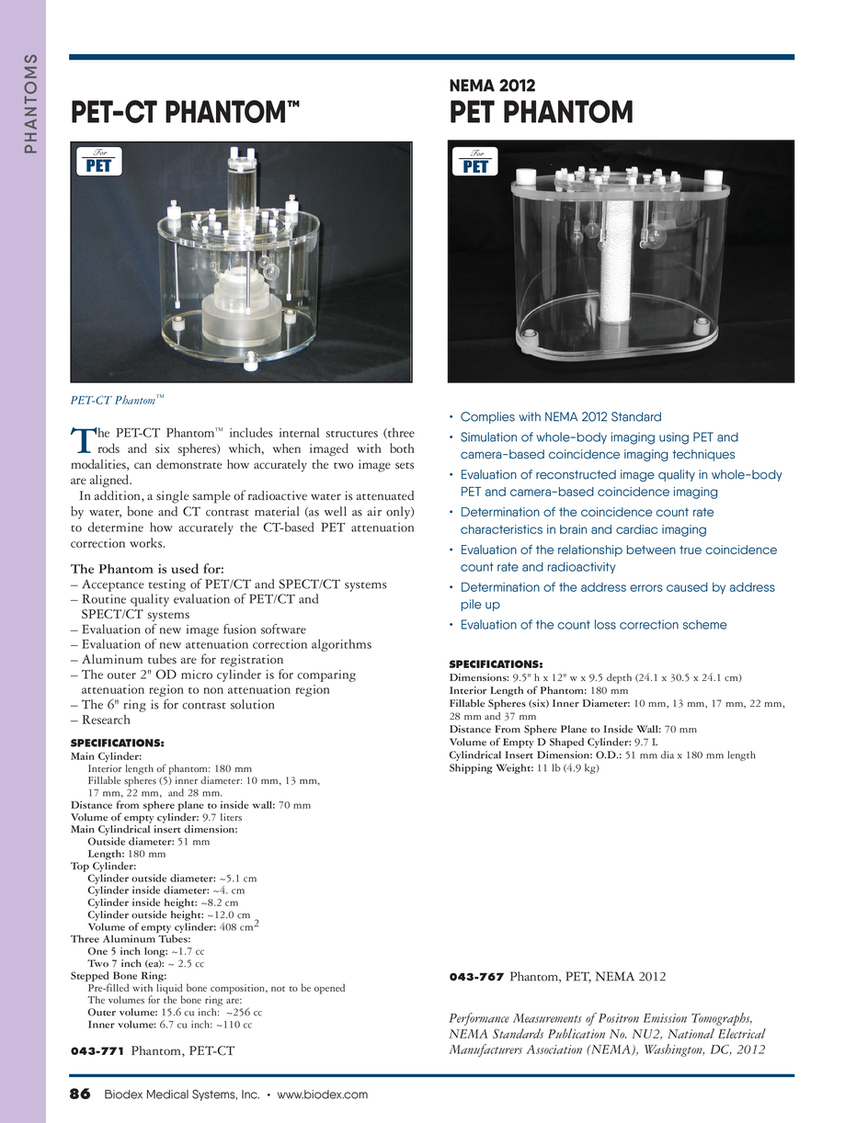
PET-CT Phantom™
T
• Complies with NEMA 2012 Standard • Simulation of whole-body imaging using PET and camera-based coincidence imaging techniques • Evaluation of reconstructed image quality in whole-body PET and camera-based coincidence imaging • Determination of the coincidence count rate characteristics in brain and cardiac imaging • Evaluation of the relationship between true coincidence count rate and radioactivity • Determination of the address errors caused by address pile up • Evaluation of the count loss correction scheme
SPECIFICATIONS:
he PET-CT Phantom™ includes internal structures (three rods and six spheres) which, when imaged with both modalities, can demonstrate how accurately the two image sets are aligned. In addition, a single sample of radioactive water is attenuated by water, bone and CT contrast material (as well as air only) to determine how accurately the CT-based PET attenuation correction works. The Phantom is used for: – Acceptance testing of PET/CT and SPECT/CT systems – Routine quality evaluation of PET/CT and SPECT/CT systems – Evaluation of new image fusion software – Evaluation of new attenuation correction algorithms – Aluminum tubes are for registration – The outer 2" OD micro cylinder is for comparing attenuation region to non attenuation region – The 6" ring is for contrast solution – Research
SPECIFICATIONS:
Main Cylinder: Interior length of phantom: 180 mm Fillable spheres (5) inner diameter: 10 mm, 13 mm, 17 mm, 22 mm, and 28 mm. Distance from sphere plane to inside wall: 70 mm Volume of empty cylinder: 9.7 liters Main Cylindrical insert dimension: Outside diameter: 51 mm Length: 180 mm Top Cylinder: Cylinder outside diameter: ~5.1 cm Cylinder inside diameter: ~4. cm Cylinder inside height: ~8.2 cm Cylinder outside height: ~12.0 cm Volume of empty cylinder: 408 cm2 Three Aluminum Tubes: One 5 inch long: ~1.7 cc Two 7 inch (ea): ~ 2.5 cc Stepped Bone Ring: Pre-filled with liquid bone composition, not to be opened The volumes for the bone ring are: Outer volume: 15.6 cu inch: ~256 cc Inner volume: 6.7 cu inch: ~110 cc
043-771
Dimensions: 9.5" h x 12" w x 9.5 depth (24.1 x 30.5 x 24.1 cm) Interior Length of Phantom: 180 mm Fillable Spheres (six) Inner Diameter: 10 mm, 13 mm, 17 mm, 22 mm, 28 mm and 37 mm Distance From Sphere Plane to Inside Wall: 70 mm Volume of Empty D Shaped Cylinder: 9.7 L Cylindrical Insert Dimension: O.D.: 51 mm dia x 180 mm length Shipping Weight: 11 lb (4.9 kg)
043-767
Phantom, PET, NEMA 2012
Phantom, PET-CT
Performance Measurements of Positron Emission Tomographs, NEMA Standards Publication No. NU2, National Electrical Manufacturers Association (NEMA), Washington, DC, 2012
86 Biodex Medical Systems, Inc. • www.biodex.com